Why Do Dentists Take a CT Scan (CBCT)? — How It Differs From a Panoramic X-ray, and Is the Radiation Safe
Dr’s Notes
“You already took a panoramic X-ray — do I really need a CT too?” It’s one of the most common questions in my operatory, and it carries two worries inside it: one about cost, one about radiation. Both deserve straight answers, so this post takes them head-on.
What's the difference between a panoramic X-ray and CBCT?
A panoramic X-ray is a two-dimensional image of your whole mouth unrolled onto one film. Every upper and lower tooth, the jawbones, the jaw joints, even the sinuses — all surveyed at a glance. As a map of your overall condition it has no equal, and for most dentistry, a panoramic plus small targeted films is entirely sufficient.
Its limit is that it’s a flattened picture. Structures sitting in front of and behind each other get stacked onto one plane, so it cannot tell you whether a wisdom tooth root passes over or behind the nerve canal, how many millimeters wide the bone is, or in which direction a lesion has spread. A map shows you where the mountain is — not its height or slope.
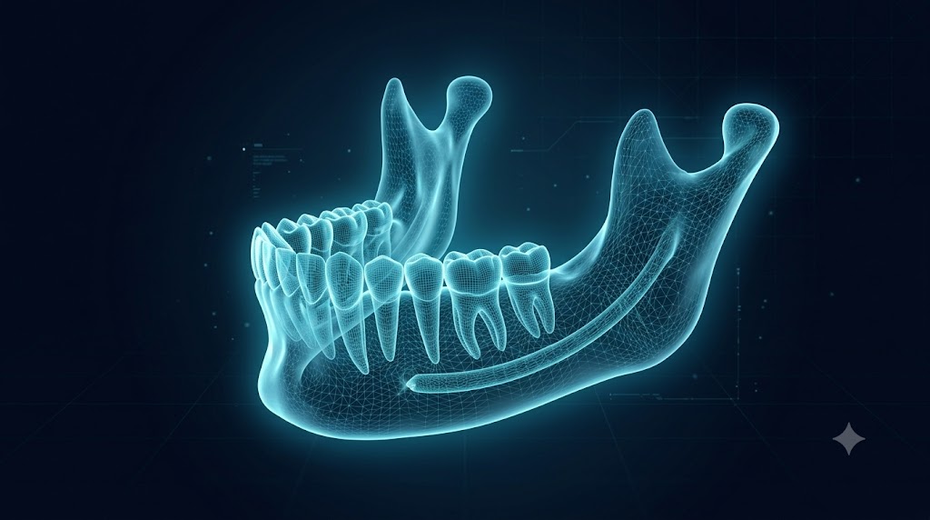
CBCT (cone-beam CT) is three-dimensional. The machine rotates once around your head, and the data is reconstructed so any spot can be sliced and viewed from any direction. The exact distance between a root and the nerve canal, the width and height of bone, the 3D extent of a lesion — CBCT answers precisely the questions a panoramic cannot.
So when is it actually needed?
Not for everyone. These are the situations where CBCT genuinely changes the quality of diagnosis and treatment:
Implant planning. Implants are millimeter-scale procedures. Placing one without confirming bone width, height, density, and the position of the nerve canal or sinus in 3D is like boring a tunnel without a survey. The decision about whether — and how much — bone grafting is needed also comes from this scan.
An impacted wisdom tooth near the nerve canal. When the panoramic shows roots overlapping the canal, CBCT reveals the true 3D relationship, which determines whether and how to extract — or whether to refer to a hospital.
Root canal failure with an unclear cause. Missed canals, suspected root cracks, the true extent of a lesion at the root tip — CBCT establishes the cause in 3D before committing to retreatment or an apicoectomy.
Other uses include evaluating bony changes in the jaw joint, assessing teeth and bone after trauma, and mapping cysts or lesions. Conversely — if CBCT is being ordered routinely for a regular checkup, a simple cavity, or a cleaning consultation, you’re entitled to ask why.
Radiation, in actual numbers
This is the bigger worry, so let’s use numbers. The effective dose of a dental CBCT varies with the field of view and machine settings, but a small-volume scan runs roughly in the range of tens to around a hundred microsieverts (μSv). For perspective: the natural background radiation we all receive from the environment is roughly 2,400–3,000 μSv per year — about 7–8 μSv per day. A small-field CBCT is therefore comparable to several days’ to a few weeks’ worth of ordinary background exposure, and it is an order of magnitude below a medical full-body CT.
That doesn’t license casual scanning. The governing principle of all radiography is to image only as much as the diagnostic question requires (the ALARA principle), and in practice it works like this: if the question can be answered in 2D, stop at the panoramic; use CBCT only when 3D information will change a treatment decision — and then only the small field that’s needed. Children are more radiosensitive than adults, so their indications are judged more strictly; during pregnancy, non-urgent imaging is deferred as a rule.
As a patient, one question before the scan is enough: “What are we trying to confirm with this image?” If a clear answer comes back — nerve distance, bone width, lesion extent — the scan has a reason to exist.
Frequently asked questions
Isn’t taking a CBCT after a panoramic redundant? They do different jobs. The panoramic is the overall map; CBCT is a precision survey of one spot. CBCT is added when something found on the panoramic needs 3D confirmation — and skipping the panoramic to shoot only CBCT would actually miss the overall picture.
Does CBCT radiation accumulate in the body? Radiation risk is a matter of probability rather than a poison that builds up, and one dental CBCT equals a few days of natural background exposure. The principle is still not to repeat unnecessary scans — and bringing your previous imaging with you is the single best way to avoid duplication.
Can children have a CBCT? Yes when genuinely needed, but the threshold is stricter than for adults, and the scan volume and settings are reduced to a child’s size. This is the group for whom “why is this necessary” deserves the clearest answer of all.
This article is for general information; individual diagnosis and treatment planning require an in-person examination.